
Educational
Ultrasound-Guided Biopsy: Minimally Invasive Precision
Dr. Francisco Delgado · August 4, 2026
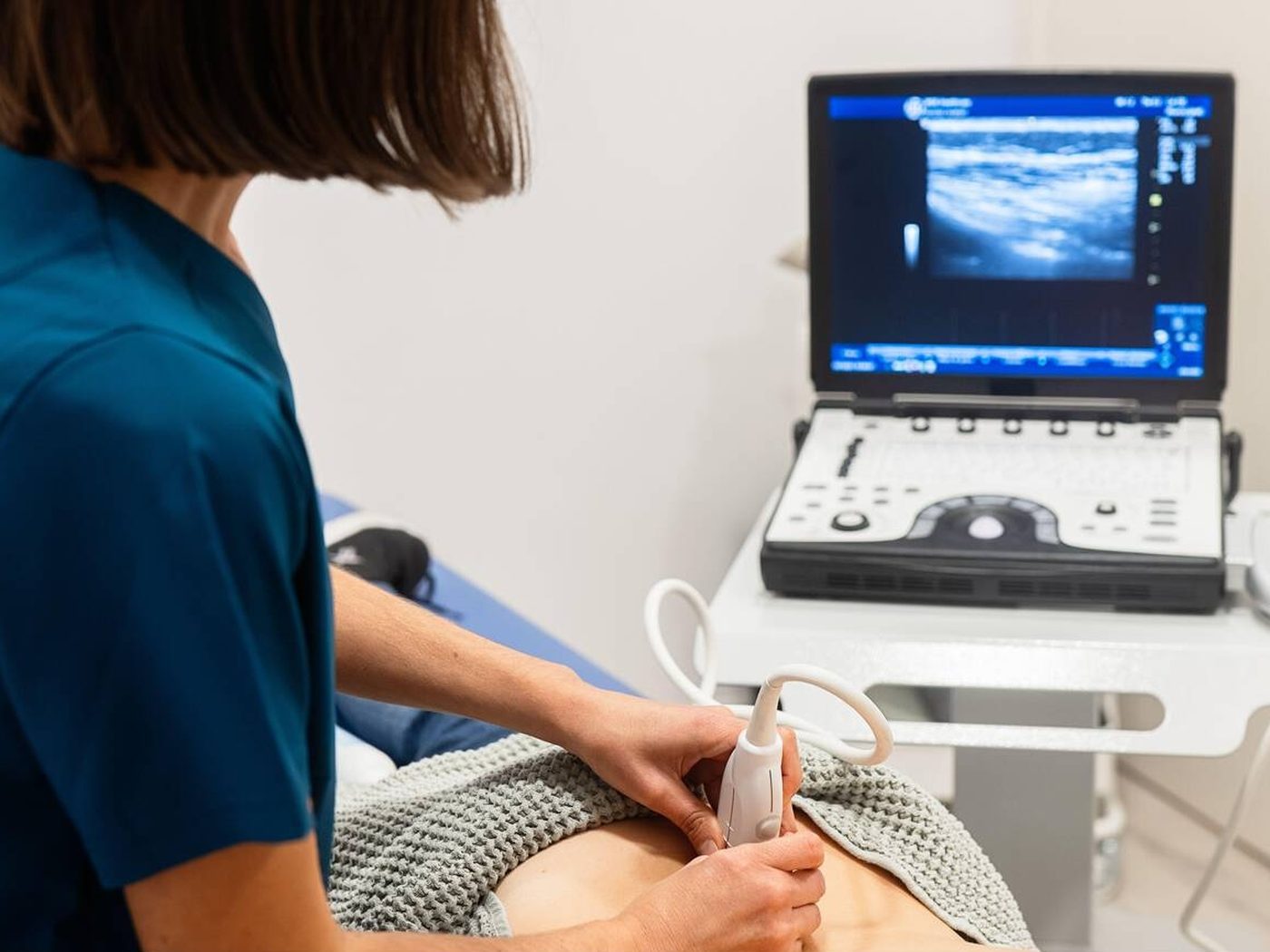
A safe outpatient diagnostic procedure guided in real time by ultrasound to obtain tissue samples with high precision — without open surgery in most cases.
When it is indicated
Ultrasound-guided biopsy is offered when a nodule or suspicious area is visible on ultrasound and the radiologist needs a histological sample to confirm or rule out malignancy (or other conditions). It is also used for axillary nodes or other ultrasound-accessible lesions, as clinically indicated.
Not every unusual image ends in biopsy. The finding is classified first (for example with BI-RADS in breast imaging) and the team decides whether follow-up, further imaging, or sampling is appropriate.
How it is performed
- Localisation: the lesion is identified in real time with the ultrasound probe.
- Local anaesthesia: the area is numbed; you remain awake and can help with positioning.
- Sampling: core-needle (or, in selected cases, aspiration) obtains tissue cores. The number of passes is set by the interventionalist.
- Quality check: the sample is confirmed as representative; a tiny metallic marker may be left to locate the site on future studies.
- Closure: local compression, dressings, and aftercare instructions.
The procedure is usually outpatient. Needle time is short once sampling starts; total room time depends on preparation and anatomy.
Recovery and aftercare
- Keep the area clean and dry as instructed.
- Bruising, tenderness, or a small haematoma for a few days is common.
- Avoid intense exercise or heavy lifting for the period advised (often 24–48 hours).
- Use your usual analgesic if your doctor allows it; do not self-medicate with anticoagulants or anti-inflammatories if they were contraindicated.
- Seek care promptly for heavy bleeding, infection signs, fever, or worsening pain.
Pathology results may take several days. Your treating physician interprets them together with imaging to define follow-up, surgery, or treatment.
Ultrasound guidance vs other methods
When the lesion is well seen on ultrasound, US guidance avoids extra radiation and shows the needle live. If the finding is only mammographic (for example a cluster of microcalcifications), stereotaxis may be preferred. MRI guidance is reserved for specific scenarios. The interventional radiology team chooses the method by lesion type — not by fashion.
At Almavita
Diagnostic intervention is part of the breast pathology and radiology pathway. If a biopsy was recommended or you want an imaging second opinion, book via WhatsApp with your recent studies.
Note: educational content. Indication, technique, and exact aftercare are defined by the specialist performing your procedure.