
Educational
Prenatal Care: The Ultrasound Schedule Every Mother Should Know
Dra. Laura Henao · August 12, 2026
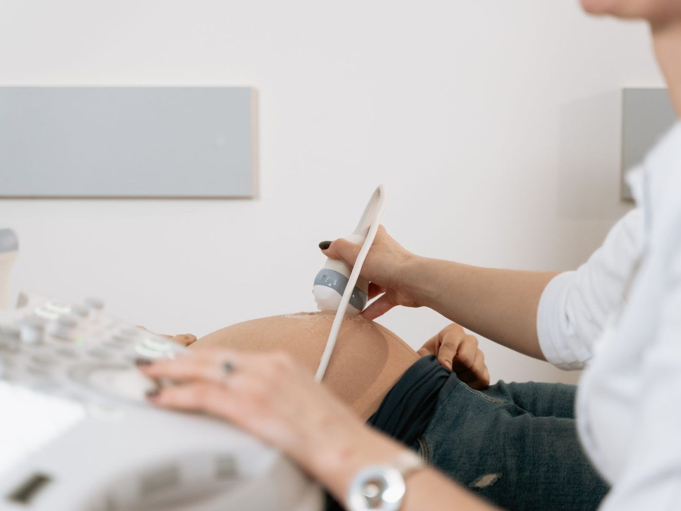
Structured ultrasound follow-up during pregnancy helps monitor maternal–fetal wellbeing and detect situations that need specialised care in time.
Prenatal care is more than “seeing the baby”
Each visit combines clinical assessment (blood pressure, weight, symptoms), trimester-appropriate labs, and ultrasounds at key moments. Ultrasound does not replace the rest of prenatal care: it adds information on gestational age, growth, anatomy, and placenta.
Exact timing is set by your obstetrician based on the course of pregnancy, prior pregnancies, and findings. What follows is an orientation map, not a rigid order.
First trimester (approx. through 13+6 weeks)
- Early ultrasound: confirms intrauterine pregnancy, viability, and number of embryos; estimates gestational age most accurately when done on time.
- Aneuploidy screening: depending on your plan (combined biochemistry, cell-free DNA, or other tests), it may coincide with a nuchal translucency scan in specific windows.
If there is bleeding, pain, or doubt about pregnancy location, do not wait for the “routine” appointment — seek care immediately.
Second trimester — the anatomy scan
Between weeks 18 and 24 (your physician sets the precise range) comes the morphological or detailed anatomy ultrasound. It reviews brain structures, face, heart, spine, abdomen, kidneys, limbs, placenta, and amniotic fluid. It is one of the most important studies of pregnancy: it is not a “4D keepsake video,” even if pleasant images are sometimes obtained — its purpose is diagnostic.
If something is incomplete (fetal position or body habitus), you may be scheduled for a complementary scan.
Third trimester
Growth, presentation, placenta, fluid, and — when indicated — Doppler are assessed. Not every pregnancy needs the same number of late scans: a low-risk pregnancy has a different calendar from gestational diabetes, hypertension, growth restriction, or multiples.
When to call outside the schedule
Reduced fetal movements (once you feel them regularly), bleeding, fluid leakage, severe headache with visual changes, epigastric pain, regular contractions before term, or fever. When in doubt, it is better to be evaluated.
Practical preparation
Ask whether you should arrive with a full bladder (more common in the first trimester). Bring your prenatal card and prior studies. Wear two-piece clothing for easier abdominal access.
At Almavita
The team’s obstetrics and gynecology practice includes obstetric ultrasound as part of accompaniment. To coordinate prenatal care or an ultrasound ordered by your physician, write us on WhatsApp.
Note: educational schedule. Your obstetrician adapts frequencies and tests to your specific pregnancy.