
News
New High-Definition Digital Mammography Technology at Almavita
August 20, 2026
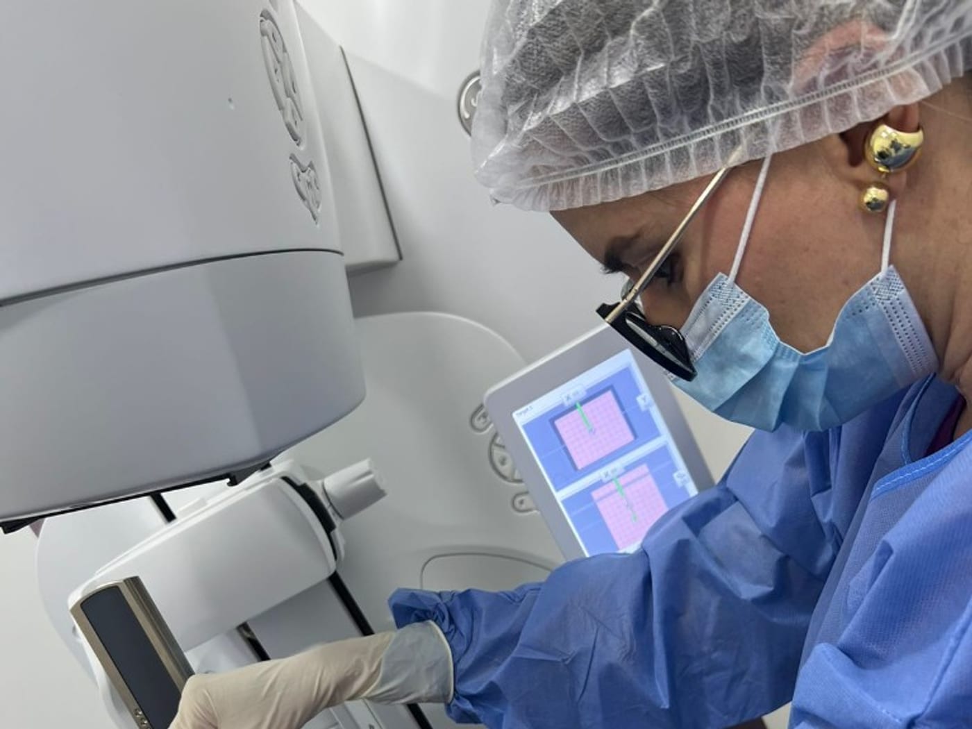
At Almavita, the breast pathology pathway relies on high-definition digital imaging and guided intervention, so each finding is seen clearly and — when needed — confirmed with the least possible invasiveness.
What digital mammography adds
Unlike older analogue systems, digital mammography captures the image electronically: contrast can be adjusted, regions of interest enlarged, and studies archived for comparison over time. That improves reading of microcalcifications and dense tissue, always within low-dose protocols and quality control.
Technology does not replace the radiologist: it strengthens judgement. A precise system poorly interpreted is useless; a precise system in expert hands shortens diagnostic pathways.
Intervention when imaging is not enough
If a finding needs histological confirmation, the team performs guided procedures. For stereotactic biopsies, the group works with high-precision platforms (including GE Pristina family systems in the team’s working environment) that localise mammographic lesions — such as microcalcification clusters — with millimetre accuracy.
When the lesion is better seen on ultrasound, ultrasound-guided biopsy is preferred. The method is chosen by the lesion, not by aesthetic preference.
What the patient gains
- Clearer images to decide follow-up or biopsy.
- Ability to compare prior digital studies.
- Outpatient procedures when sampling is indicated.
- One clinical team that knows the breast history end to end.
How to access care
If your insurer or treating physician ordered a mammogram, breast ultrasound, or an interventional procedure, contact us on WhatsApp. Bring prior studies: comparison is part of diagnostic value.
Note: informational note on diagnostic capabilities. Indication for each study is set by your physician.